|
|
||
 |
November 13, 2006 - SLAC Researchers Help Demonstrate Potential of FEL Imaging - Press Release
-- return to Press Releases --
Date Issued: November 13, 2006 Contact:
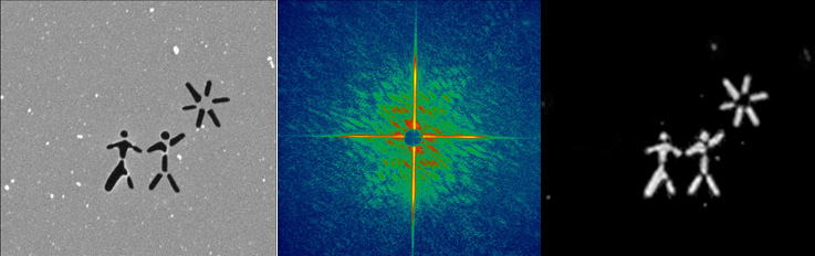
Menlo Park, CA—A collaboration of scientists including researchers from the Department of Energy's Stanford Linear Accelerator Center (SLAC) have for the first time successfully demonstrated the use of extremely short and intense soft X-ray pulses to capture images of objects before the pulses destroy the sample. The team also set a speed record of 25 billionths of a second of the duration of the X-ray pulse used to acquire the image. The results will be published in the November 12 online edition and the December print edition of Nature Physics. Called "flash imaging," this experiment proves the principle behind atomic-scale imaging of complex biomolecules that will be applied when even more powerful X-ray lasers are available, such as the Linac Coherent Light Source (LCLS), now under construction at SLAC; the SPring-8 Compact SASE Source (SCSS) facility in Japan; and the European XFEL in Hamburg. This revolutionary tool will give scientists unprecedented insight into fields such as materials science, chemistry and biology and medicine. "This result is a remarkable validation of the concept of imaging using single pulses from a free electron laser," said Keith Hodgson, Director of Photon Science at SLAC and a co-author of the paper. "This is just the first glimpse of the breakthrough discoveries that will come from LCLS when it becomes operational in 2009." Using the free-electron laser at Deutsches Elektronen-Synchrotron (DESY) in Hamburg, an international collaboration led Henry Chapman of Lawrence Livermore National Laboratory (LLNL) and Janos Hajdu of SLAC and Uppsala University zapped a sample containing nanometer-sized objects and recorded the pattern of scattered X-rays—the diffraction pattern—before the laser destroyed the sample. A special computer algorithm was then used to recreate an image of the object based on the recorded diffraction pattern. Computer simulations suggest that near-atomic resolution could be achieved by well-thought out choice of pulse length and intensity of X-ray wavelength before the sample is stripped of its electrons and destroyed. However, up until now, there had been no experimental verification of the technique. A theory proposed by Hajdu predicts that a single diffraction pattern may be obtained from a much smaller object, such as a macromolecule, virus or cell, by using an ultra-short and extremely bright X-ray pulse before the sample explodes and turns into a plasma. Successfully demonstrating the workability of this theory using soft X-rays means that scientists will soon be able to apply this technique using X-rays of a much shorter wavelength to study much smaller objects. These "hard" X-rays, such as will be first produced by the LCLS, will ultimately enable the study of macromolecular proteins without first having to grow them into crystals. Because many biomolecules resist crystallization, flash imaging will finally enable the rapid study of all classes of proteins. Scientists from Lawrence Livermore National Laboratory, Uppsala University in Sweden, SLAC, DESY, Technische Universität Berlin, the Center for Biophotonics Science and Technology at U.C. Davis, and the private firm Spiller X-ray Optics of Livermore, conducted the first experimental demonstration of this theory. This experiment was conducted using the first soft X-ray free-electron laser in the world at the FLASH facility at DESY. In addition, this work showed that it takes a pulse of only 25 femtoseconds to capture an image. Until now, there had been uncertainty as to whether a diffraction pattern could be recorded under these circumstances and subsequently reconstructed to obtain an image of a sample before it was damaged. "The entire collaboration is very excited by these results," said Hajdu. "Flash imaging has implications for studying molecular structures in biology in a whole new way. A new scientific community is forming to achieve these goals by combining biology with atomic, plasma, and astrophysics for the first time." The work was funded in part by the U.S. Department of Energy Office of Science and by a Laboratory Directed Research and Development strategic initiative proposal for "biological imaging with fourth-generation light sources" at LLNL. by Brad Plummer -30- |
Last update: